

Nanoscope Systems, Inc
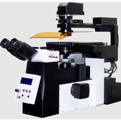
Model ABM -Automatic Biological Microscope
ABM is a classical type inverted automatic biological microscope for fluorescence imaging. Also with the observation through microscope eyepieces, all the microscope images are displayed on a monitor by the camera attached to the side-port of the microscope body, and saved as electric image files of standard formats. Fully automated stages and revolving turret are controlled by the operation software. Convenient microscope observation with ABM will maximize the efficiency of your microscope experiments.
Most popular related searches
biological microscope
well plate
microscopy
fluorescence imaging
automatic microscopy
fluorescence microscope
bio-imaging
objective lens
petri dish
inverted microscope
- Inverted fluorescence microscope
- 5 color fluorescence imaging
- DIC (differential interference contrast) imaging
- Phase contrast imaging
- Bright field imaging
- Observation through eyepieces
- Automated X,Y,Z Stage
- Automated objective lens changer
- Automated filter wheel changer
- Slide glass, well plate, Petri dish compatible
Automatic well plate scanning is a kind of large area stitching efficiently set for the well plate format. It numbers wells automatically, and scans the circular well-areas only. Any standard well plates can be conveniently scanned.
The consecutive imaging of pre-defined area, and stitching of these acquired images will provide a lot of convenience for the wide range imaging of large samples. The stitched image can be analyzed as one single image, and/or the user can also observe original individual images to see specific segmentations of the stitched image in detail. Slide glass imaging, or well-plate scanning is available with this function.
Z-stacking (also known as focus stacking) is a digital image processing method which combines multiple images taken at different focal distances to provide a composite image with a greater depth of field (i.e. the thickness of the plane of focus) than any of the individual source images. Z-stacked images can be reconstructed as 3D images by deconvolution algorithm.