CrestOptics S.p.A.
  1. Companies
  2. CrestOptics S.p.A.
  3. Products
  4. CrestOptics DeepSIM - Super-Resolution ...
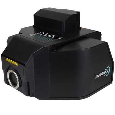
CrestOptics DeepSIMSuper-Resolution Microscopy Module

SHARE

The DeepSIM module is an innovative Lattice Structured Illumination Microscopy (SIM) tool that enhances the capabilities of traditional microscopes by providing super-resolution imaging down to 100 nm lateral and 300 nm axial resolutions. Designed to integrate with any existing microscope, it bridges the gap between widefield imaging and confocal microscopy, offering deep Z-depth penetration and high-resolution data acquisition across a variety of specimen types. Leveraging 2D Lattice SIM technology, it offers minimal phototoxicity and robust image reconstruction, making it suitable for live-cell imaging. The system supports a wide range of fluorophores and objectives, from low to high magnification, and can be used with various immersion media, allowing for versatile applications in biological research. DeepSIM is particularly beneficial for studies involving complex organisms like 3D organoids and small animal models, where it ensures high fidelity and reduced artifacts in image capture.

Most popular related searches

At CrestOptics, we believe that super-resolution should be accessible for all scientists to progress their research.
This is the reason behind the development of the DeepSIM, the first Lattice SIM super-resolution module that is compatible with any existing upright or inverted microscope with a  camera port. 

The DeepSIM constitutes the seamless evolution of any diffraction-limited microscopy approach.
It is as easy to use as a widefield system and reaches the same Z-depth penetration of a confocal microscope, enabling scientists to access super-resolved data through conventionally prepared, deep, thick specimens, even in challenging sampling conditions.

Imaging Across Scale: Multiple Lattice SIM Illumination Patterns

The 2D Lattice SIM technology behind DeepSIM enhances the illumination efficiency and homogeneity with minimal phototoxicity in live cell imaging experiments. Besides, the 2D Lattice SIM Illumination delivers a better contrast that significantly improves in-depth Z-acquisitions and the image reconstruction robustness

More importantly, the user can choose among 3 Lattice SIM Illumination Patterns for approaching specimens of increasing thickness and morphological complexity. The well-matched sampling conditions significantly mitigate the risk of artifacts in image reconstruction.

High throughput

  • Live imaging
  • Cell monolayers
  • Thin samples (up to 50 µm)
  • Dotted or filiformed structure

Standard

  • 3D cell culture
  • Uncleared tissue
  • Thick samples up to 100 µm
  • Intricate structures (e.g. glomeruli) 

Deep Imaging

  • Small organisms (C. elegans, Zebrafish)
  • Cleared tissue
  • Deep imaging over 100 µm
  • Dense and homogeneous structures

Down to 100 nm lateral resolution

Featuring the structured lattice illumination with the computationally super-resolved data reconstruction, DeepSIM improves the spatial resolution in all three dimensions reaching 100 nm laterally and 300 nm axially.

HeLa cells with Oregon Green™ 488 stained tubulin. Two progressive zooms of the area of interest (dotted box) are shown in the upper right boxes. The intensity profile obtained on the tubulin filament under the red arrow is depicted in the graph. 100x Oil, NA 1.45.

Deep imaging through thick specimens

Deep super-resolved data across conventionally prepared thick samples. The DeepSIM is designed to work with samples of thicknesses comparable to those used with Confocal microscopy, giving super-resolved data over 50 μm Z-depth. Z-stacks take full advantage of the entire objective working distance and meaningful data can be obtained from a native heterogeneous and intricate sample, providing researchers with a complete understanding of its structure and composition. 

Cleared mouse brain with GFP-neurons is shown in 3D view depth-code (130 µm). 60x Oil, NA 1.4. 

Suitable with standard confocal staining up to Near-Infrared 750 nm Excitation

Multiplexed imaging is an emerging way to gather information from multiple cellular markers simultaneously. DeepSIM performs optimally with the standard staining markers for confocal microscopy. It is the only SIM technology covering the full spectrum from 405 nm to NIR 750 nm excitation wavelength. DeepSIM offers the maximum flexibility in fluorophore choice and reduces the spectral overlap in multicolor experiments. 

3D movie of a mouse brain tissue section showing neurons with dendritic spines (yellow), glial cells (magenta), astrocytes (white) and DNA (cyan). 60x oil 1.4 NA. Sample credits: Prof. Di Angelantonio, CLN2S@Sapienza Università di Roma – IIT.

Compatible with Low Mag objectives and with any kind of Immersion Media

The DeepSIM was conceived to maintain high standards of performance over a large set of objectives from Low Magnification 20x up to High Magnification 100x. The compatibility with dry lenses and any immersion media expands the range of suitable applications over a large cohort of sample and support types. DeepSIM offers the best trade-off between FOV and resolution to catch fine details in extra-large objects.

(i) Mouse retina section stained with nuclei (blue), Vimentin (green), CD39 (orange), nuclear marker (red). A Maximum Intensity Projection (MIP) of 100 µm Z-stack is shown. 25x Silicone, NA 1.05. Sample credits: Dr. Lagerholm, Dr. Yegutkin, Dr. Svärd (University of Turku&InFLAMES Research Flagship, Finland), and Dr. Imreh (Karolinska Institutet, Sweden). 

Click here to see the 3D-movie.

(ii) DeepSIM acquisition of the Zebrafish eye region with Cy7 neuronal marker. A MIP of 70 um Z-stack is shown. 20x Dry, NA 0.8. Sample credits: Dr. Paradisi, Dr. Tartaglia, Dr. Lauri, Ospedale Pediatrico Bambino Gesù, Rome, Italy.

Resilient resolving power across specimen scale: from 3D organoids to small animal models

The DeepSIM resolving power is preserved across a wide spatial scale, including 3D organoids, tissue slices, and small animal models (Zebrafish, C. elegans). The extended flexibility suggests its use with Spinning Disk technology in a correlative bioimaging approach.


(i) Dissociated neurons co-cultured with cancer organoids: a MIP of a 13 μm Z-stack is shown. Staining: nuclei with DAPI (blue), EpCAM for cancer cells (green), b3 Tubulin (red), SV2A (synaptic density marker, orange), and Synaptophysin (neuroendocrine presynaptic marker, white). 60x Oil, NA 1.42. Sample credits: Prof. Hwang, Dr. Wang, Harvard University, MA, USA 

(ii) Caenorhabditis elegans embryo stained with DAPI (blue) and Alexa568 showing protein condensates formed by the transcription factor HSF-1(orange). A MIP of a DeepSIM acquisition is shown. 100x Oil, NA 1.45. Sample credits: Prof. Klosin, Nencki Institute of Experimental Biology PAS, Warsaw, Poland

(iii) A MIP of a 40  μm  Z-stack of a whole mouse brain slice with GFAP-positive astrocytes. 60x Oil, NA 1.42. Sample credits: Dr. Brunne and Dr. Failla, Universitätsklinikum Hamburg Eppendorf UKE – Microscopy Imaging Facility, Germany.