


- Home
- Companies
- Tomocube Inc.
- Products
- Tomocube - Model HT-X1 - Live-Cell ...
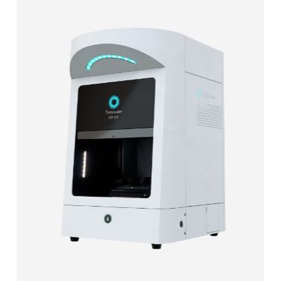
Tomocube - Model HT-X1 -Live-Cell Imaging System
The Tomocube HT-X1 system is a pivotal innovation in live-cell imaging, utilizing Holotomography technology to allow for high-resolution, three-dimensional observation of cellular structures without the need for labeling or fixation. It employs low-coherence light sources which enhance the quality of imaging by reducing phototoxicity and noise, making it especially suitable for delicate samples such as stem cells and organoids. The HT-X1 is equipped with a versatile illumination module integrated with a Digital Micro-mirror Device, enabling rapid cycling through illumination patterns to achieve a high numerical aperture, essential for high-content imaging. Additionally, its compatibility with multiple vessel types, like 96-well plates, and the incorporation of TomoAnalysis software for quantitative analysis, make it a versatile tool in cellular research. Its real-time monitoring capabilities and integrated environmental control further facilitate extensive and precise cellular study.
The #1 Choice for Live-Cell Imaging and Analysis Empowering your Exploration. Redefining the Limits.
Holotomography is a technique that utilizes low-intensity light to acquire the refractive index of cells from multiple angles. This technology has emerged as an excellent cellular imaging solution, allowing for high-resolution imaging while maintaining the health of the cells by using low-intensity light sources. Tomocube's second-generation Holotomography, the Tomocube HT-X1, employs low-coherence light sources, ensuring lower toxicity and noise-free high-resolution image acquisition compared to laser-based methods.
The high-resolution images obtained through this technology enable real-time observation of not only the morphology of living cells but also the shapes of subcellular organelles, such as the nucleus, nucleolus, mitochondria, and lipid droplets. The integrated stage-top incubator provides a stable cultivation environment, enabling prolonged monitoring of sensitive cells like stem cells or organoids.
This innovative system is compatible with various commercial imaging plates and supports imaging up to 96-well plates, making it applicable for high-throughput screening. Utilizing the HT-X1 allows for the acquisition of high-resolution images of cells, microorganisms, organoids, and tissue samples. Additionally, Holotomography analysis software, TomoAnalysis, facilitates obtaining quantitative information based on refractive index, including high-resolution 3D images of cells, as well as volume, area, concentration, and dry mass. Therefore, the use of HT-X1 becomes a crucial tool in understanding biological phenomena.
Label-free 3D visualization
Label-free 3D live cell imaging of monolayered cells and 3D organoids
Correlative fluorescence
Correlative fluorescence imaging to jointly obtain biomolecular specificity information
Long-term timelapse
Built-in incubator that provides a stable cell culture environment
High-throughput screening
Multi-well plate compatibility for high-throughput experiment
Quantitative analysis
Quantitative measurements of cells and subcellular components
High-Resolution Three-Dimensional Imaging
HT-X1 uses an LED illumination module integrated with a Digital Micro-mirror Device (DMD). When HT-X1 captures a 3D image, the illumination module is rapidly cycling through a set of optimized illumination patterns programmed in the DMD to encode specific spatial features of the specimen. By synthesizing these images acquired from multiple angles, HT can overcome the optical resolution limit, achieving higher synthetic NA compared to the physical NA value of objective lens. Therefore, HT-X1 imaging provides high-content, high-resolution 3D imaging at exceptional stability.
Label-free Observation of Cellular Dynamics
The Holotomography approach in live cell imaging does not require fixation or labeling, which reduces unwanted artificial intervention and the need for long exposure to light. By minimizing the risks of phototoxicity and photobleaching, the specimen remains at its nature state for observation. With a rapid capture speed of 1 second per frame, HT enables real-time monitoring of dynamic cellular processes with exceptional temporal resolution. Additionally, its integrated stage-top feature facilitates long-term observations, empowering researchers to explore cellular phenomena over extended periods with stability and precision.
Versatile Imaging Capability for Multi-Well Plates
Especially designed to maximize user flexibility, the HT-X1 employs a unique adaptive illumination module that is tailored for multi-well plates. The combination of high NA, a long working distance condenser, DMD, and a motorized illumination unit delivers an efficient illumination pattern for a diverse range of vessel types, from 35-mm dishes to 96-well plates. With the HT-X1, researchers will have more freedom to design their experiments on different sample types using any imaging vessel of their choice.
Intuitive Software with Enhanced Control
TomoStudio X works in concert with the HT-X1 platform to visualize and analyze RI tomograms. This intuitive, easy-to-use software empowers users with complete system control, allowing them to handle even the most complex experiments through simple mouse clicks. Researchers can easily set up complex time-lapse sequences, perform large-area tile imaging and stitching tasks, and image multiple points with ease using this software.