
Refine by
Tissue Imaging Articles & Analysis
42 news found

We are proud to introduce Myelotracker®, a peptide-based probe. This unique technology specifically targets lactoferrin, a major protein stored in neutrophil granules, making it possible to visualize and study these immune cells with unmatched precision. Unlike many existing tools, Myelotracker does not activate neutrophils, preserving their natural physiological state and ensuring reliable ...
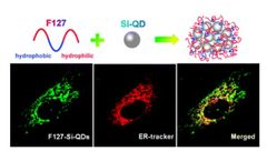
These semiconductor nanocrystals, ranging from 2-10 nm in size, emit precise wavelengths of light when excited, making them ideal for high-resolution displays, solar cells, and biomedical imaging. Alfa Chemistry’s cadmium-free QDs, such as indium phosphide (InP) and silicon-based variants, are particularly sought after for their eco-friendly profile and superior performance ...

These highly purified nanoparticles offer exceptional performance and reliability in various research and commercial applications, such as drug delivery, nanosensors, photothermal therapy, antimicrobial agents, biomedical imaging, catalysis, and tissue engineering. Silver nanoparticles (AgNPs) are increasingly being used in various fields due to their unique ...

Yang, L., et al (2024) introduces LungVis 1.0, an AI-driven imaging platform integrating light sheet fluorescence microscopy (LSFM) and deep learning for mapping nanoparticle (NP) deposition across murine lungs. The research highlights the advantages of aerosol-based delivery methods over traditional liquid-based approaches, emphasizing the uniformity of NP deposition in distal alveolar ...

CD BioSciences, a US-based biotechnology company focusing on the development of imaging technology, announced the launch of its new 3D Tissue Imaging Services to researchers with comprehensive solutions for 3D imaging of tissues, including 3D Immunolabeling, Tissue Removal, 3D ...
Compared with traditional imaging and spectroscopy, image processing and analysis are the core of hyperspectral imaging technology. ...

” Jeremy Sobotta, Perimeter’s Chief Executive Officer commented, “This publication further supports the potential use of our flagship Perimeter S-Series OCT medical imaging technology across several tissue types, including head and neck surgery. ...
Perimeter S-Series OCT received FDA 510(k) clearance in 2021 and is a medical imaging tool that uses Optical Coherence Tomography (OCT) to provide clinicians with cross-sectional, real-time margin visualization (1-2 mm below the surface) of an excised tissue specimen. ...

As part of this grant, breast surgeon collaborators will use OnLume’s imaging device for intraoperative visualization and assessment of small lymphatic tissue and blood vessels. The FGS imaging system may help reduce the risk of two breast cancer-related morbidities, lymphedema and tissue necrosis. ...

Short for wide-area transepithelial sampling with three-dimensional analysis, WATS3D utilizes enhanced tissue acquisition, 3D imaging with AI-powered analysis, and expert pathologists to reliably detect Barrett’s esophagus (BE) and esophageal dysplasia. ...

Broncholab provides physicians with reproducible CT values for pulmonary tissue for providing quantitative support for diagnosis and follow-up examination. Broncholab can be used to support physicians in the diagnosis and documentation of pulmonary tissues images (e.g. abnormalities) from CT thoracic datasets. 3D segmentation and isolation of ...
ByFluidda

AVB-620 has completed a Phase Ib clinical trial assessing safety, pharmacokinetics, and fluorescence properties using tissue image analysis. A Phase II clinical trial for AVB-620 in breast cancer is ongoing. ...
This platform helps clinicians overcome the well-known limitations associated with traditional upper endoscopic methods of screening and surveillance for BE. WATS3D utilizes advanced tissue sampling, 3D imaging technology and an Artificial Intelligence neural network to more accurately and reliably identify BE and dysplasia. ...
Perimeter S-Series is a novel medical imaging system that uses Optical Coherence Tomography (OCT) to provide clinicians with cross-sectional, real-time margin visualization (1-2 mm below the surface) of an excised tissue specimen across a variety of tissue types. ...

Combining engineering and medicine can lead to world-leading tools like the Raman spectroscopy, which can help to distinguish cancer cells from healthy cells; for example, in brain tumour surgery.ISTOCKPHOTO / GETTY IMAGES One of the biggest challenges in brain tumour surgery is making sure that all cancer cells are removed. ...
In a recent article, Prof Daniel Salerno, Prof Victor Kim and colleagues used Functional Respiratory Imaging to better understand the crucial link between DLCO and Pulmonary Vascular Abnormalities in COVID19. ...
ByFluidda

The lipid composition varies from tissue to tissue, even within different cells of the tissue. “MALDI-Imaging is a well-known solution provided by Creative Proteomics. ...

RIVANNA, developers of imaging-based medical solutions, announced that they have received a contract from the Biomedical Advanced Research and Development Authority (BARDA), a division of the Office of the Assistant Secretary for Preparedness and Response (ASPR) within the U.S. ...

SANTA CLARA, Calif., Oct. 12, 2021 /PRNewswire/ -- Invenio Imaging is proud to announce the 2000th case with the NIO Laser Imaging System, which uses Stimulated Raman Histology to provide microscopic images of fresh tissue specimens in minutes. ...
SANTA CLARA, Calif., Sept. 28, 2021 /PRNewswire/ -- Invenio Imaging announced today that it has obtained the CE Mark for the NIO Laser Imaging System according to the In Vitro Diagnostic Directive (IVDD). ...